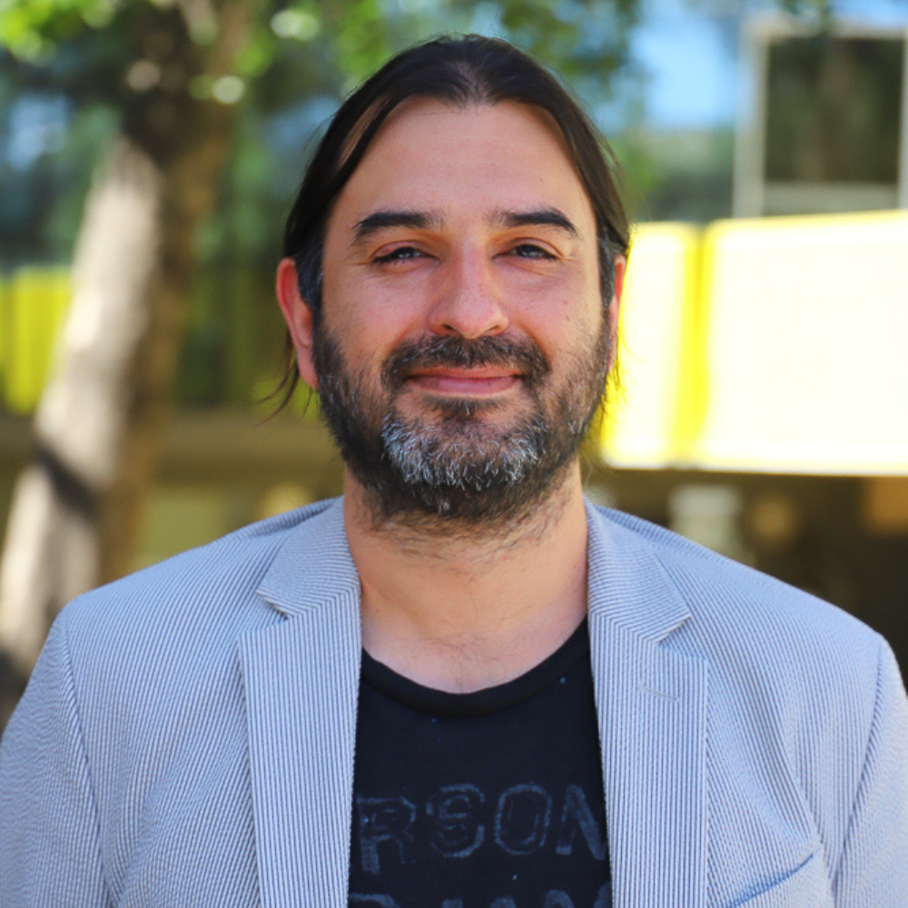
David Espíndola Rojas Profesor Asociado

Grado Académico

Doctorado en Ciencias con Mención en Física, Universidad de Santiago de Chile

Título(s) Profesional

Licenciado Física Aplicada, Universidad de Santiago de Chile

Descripción

Dr. David Espíndola received his Doctoral degree in physics from the Universidad de Santiago de Chile, Santiago, Chile, in 2012. As part of his Ph.D. dissertation, he studied the interaction wave- particle in granular materials. He pursued post-doctoral research at the Institut d’Alembert, Sorbonne Université, Paris, France, where he started conducting research on medical ultrasound. He also held a post-doctoral position with The University of North Carolina at Chapel Hill, Chapel Hill, NC, USA, where he also was a Research Assistant Professor. He is currently an Assistant Professor at the Instituto de Ciencias de la Ingeniería at the Universidad de O'Higgins in Chile. His research interests are the linear and nonlinear elastic wave propagation in soft materials, the ultrasound super-resolution imaging and the elasto-acoustics in complex medium.

15

7

  • REVISTA IEEE Access
  • 2022

Characterization of Direct Localization Algorithms for Ultrasound Super-Resolution Imaging in a Multibubble Environment: A Numerical and Experimental Study


• Aline Xavier • Héctor Alarcón • David Espíndola •

http://dx.doi.org/10.1109/ACCESS.2022.3173308

  • REVISTA 2021 IEEE UFFC Latin America Ultrasonics Symposium (LAUS)
  • 2021

Comparison of localization methods in super-resolution imaging


• David Espíndola •

http://dx.doi.org/10.1109/LAUS53676.2021.9639191

  • REVISTA IEEE Transactions on Ultrasonics, Ferroelectrics, and Frequency Control
  • 2021

Modeling ultrasound propagation in the moving brain: applications to shear shock waves and traumatic brain injury


• Sandhya Chandrasekaran • Bharat B. Tripathi • David Espíndola • Gianmarco F. Pinton •

http://dx.doi.org/10.1109/tuffc.2020.3022567

  • REVISTA Biomedical Physics & Engineering Express
  • 2020

Quantitative sub-resolution blood velocity estimation using ultrasound localization microscopy ex-vivo and in-vivo


• David Espíndola • Ryan DeRutier • Francisco Santibanez • Paul A Dayton • Gianmarco Pinton

http://dx.doi.org/10.1088/2057-1976/ab7f26

  • REVISTA International Journal for Numerical Methods in Biomedical Engineering
  • 2019

Piecewise parabolic method for propagation of shear shock waves in relaxing soft solids: One-dimensional case


• Bharat Tripathi • David Espíndola • Gianmarco Pinton •

http://dx.doi.org/10.1002/cnm.3187

  • REVISTA Journal of Computational Physics
  • 2019

Modeling and simulations of two dimensional propagation of shear shock waves in relaxing soft solids


• David Espíndola • Bharat Tripathi • Gianmarco Pinton •

http://dx.doi.org/10.1016/j.jcp.2019.06.014

  • REVISTA IEEE Transactions on Ultrasonics, Ferroelectrics, and Frequency Control
  • 2019

Super-Resolution Imaging Through the Human Skull


• Danai E. Soulioti • David Espíndola • Paul A. Dayton • Gianmarco F. Pinton •

http://dx.doi.org/10.1109/tuffc.2019.2937733

  • REVISTA Physical Review Applied
  • 2018

Focusing of Shear Shock Waves


• David Espíndola • Bruno Giammarinaro • Francoise Couvlouvrat • Gianmarco Pinton •

http://dx.doi.org/10.1103/PhysRevApplied.9.014011

  • REVISTA Physical Review E
  • 2018

Amplification of stick-slip events through lubricated contacts in consolidated granular media


• David Espíndola • Belfor Galaz • Francisco Melo Hurtado •

http://dx.doi.org/10.1103/PhysRevE.98.042907

  • REVISTA Shock Waves
  • 2017

Piecewise parabolic method for simulating one-dimensional shear shock wave propagation in tissue-mimicking phantoms


• David Espíndola • Bharat B. Tripathi • Gianmarco Pinton •

http://dx.doi.org/10.1007/s00193-017-0734-8

  • REVISTA Physical Review Applied
  • 2017

Shear Shock Waves Observed in the Brain


• David Espíndola • Stephen Lee • Gianmarco Pinton •

http://dx.doi.org/10.1103/PhysRevApplied.8.044024

  • REVISTA Theranostics
  • 2017

3-D Ultrasound Localization Microscopy for Identifying Microvascular Morphology Features of Tumor Angiogenesis at a Resolution Beyond the Diffraction Limit of Conventional Ultrasound


• David Espíndola • Fanglue Lin • Sarah E. Shelton • Juan D. Rojas • Gianmarco Pinton

http://dx.doi.org/10.7150/thno.16899

  • REVISTA Physical Review E
  • 2016

Creep of sound paths in consolidated granular material detected through coda wave interferometry


• David Espíndola • Belfor Galaz • Francisco Melo Hurtado •

http://dx.doi.org/10.1103/PhysRevE.94.012901

  • REVISTA Physical Review E
  • 2013

Effect of packing fraction on shear band formation in a granular material forced by a penetrometer


• David Espíndola • Franco Tapia • Eugenio Hamm • Francisco Melo Hurtado •

http://dx.doi.org/10.1103/PhysRevE.87.014201

  • REVISTA Physical Review Letters
  • 2012

Ultrasound induces aging in granular materials


• David Espíndola • Belfor Galaz • Francisco Melo Hurtado •

http://dx.doi.org/10.1103/PhysRevLett.109.158301

  • Enero 2022
  • - Enero 2026
Proyecto Adjudicado

During the last decades, compelling evidence shows how the context in which early life takes place impinges risk or protection for later development of non-communicable chronic diseases. In this regard, impaired fetal growth, as occur in the fetal growth restriction (FGR), leads to a higher risk for later cardiovascular diseases, an effect that would be mediated by accelerated aging at molecular, structural, and functional levels. FGR remains a leading cause of perinatal morbidity and mortality, affecting ~10% of pregnancies, but ranging 5 to 25% depending on the nutritional and health conditions of the population surveyed, with a higher prevalence among pregnant women of low socioeconomic status. In the clinic, FGR is normally defined by a fetal weight below the 10th percentile, however, new evidence shows that impaired intrauterine growth may affect several neonates born over the 10th percentile, which may be missed from the perinatal survey for preventing adverse outcomes. This points out the need for further studies to improve the understanding and identification of altered fetal growth trajectories and their consequences on vascular function. Studies in placenta show that FGR vascular dysfunction is also found at birth in chorionic and umbilical arteries. We have demonstrated the presence of functional and molecular markers (e.g. epigenetic changes) of endothelial dysfunction in human umbilical and chorionic vessels, findings that have been further confirmed by comparing systemic (aorta and femoral arteries) and umbilical arteries in animal models of FGR. These traits suggest that umbilical artery endothelial cells (HUAEC) can be used as a surrogate to explore the vascular programming within the fetus, however, their translation to clinical preventive applications for promoting healthy aging deserves further studies. It worth noting that fetal reduced oxygen supply (i.e. fetal hypoxia) and altered blood flow patterns (i.e. shear stress) are key clinical markers in the FGR, independently of the constraints leading to impaired growth, and both factors exert a tight control of vascular development and function across life. However, how these key stimuli interact and impose an epigenetic program on the endothelial function remains elusive. This proposal will focus on the crosstalk between hypoxia and shear stress that results in the endothelial programming related to impaired fetal growth, and the molecular mechanisms that mediate the vascular responses to these stimuli. Furthermore, we will address if these molecular markers may allow detecting early vascular aging in FGR subjects beyond the 10th centile cutoff. We hypothesize that “Impaired fetal growth conditions are associated with epigenetic programming of aging- and mechanosensing-related miRNAs and transcripts in the endothelium, which can be triggered by the confluence of altered flow patterns and hypoxia resulting in molecular and structural pro-hypertensive biomechanical vascular properties”. This hypothesis will be addressed by three General Objectives (GO) involving ex vivo, in vitro, and in vivo observational and mechanistic approaches: GO1 To demonstrate, in HUAEC, whether late FGR results in epigenetic changes related to the regulation of vascular aging and the expression of mechanosensing mechanisms involved in the endothelial-dependent relaxation, and their relationship with general prenatal parameters of vascular health. GO1 will be performed by recruiting HUAEC samples from late FGR and control pregnancies, to assess transcriptomic and DNA methylation analyses that will be crossed with prenatal clinical data. GO2 To study, in vivo, whether stimuli related to FGR (i.e. hypoxia and altered shear stress) differentially regulate mechanosensing pathways involved in the endothelial-dependent relaxation and their relationship with the in vivo and ex vivo vascular properties (e.g. functional and biomechanical). GO2 will be performed in chicken embryos exposed to hypoxia and treated with agents targeting mechanosensing pathways, in which wall shear stress will be determined by Ultrasound Localization Microscopy, with complementary functional, structural, and molecular analyses. GO 3. To study, in cultured HUAEC, whether stimuli related to impaired fetal growth converge in the regulation of mechanosensing-and aging-related transcripts and miRNA, contributing to the cellular programming of endothelial dysfunction. OG3 will be performed in HUAEC exposed, in vitro, to sustained hypoxia and diverse flow patterns (shear stress), in which target DNA methylation, miRNA, transcripts, and proteins will be assessed. Our expected outcome is to improve the knowledge about the endothelial epigenetic programming after FGR and enhance the characterization of in vivo shear stress patterns and mechanisms induced by chronic fetal hypoxia. This project is not only relevant to uncover the developmental approaches for diagnosing and treatments in complicated pregnancies.
Co-Investigador/a
  • Diciembre 2021
  • - Noviembre 2023
Proyecto Adjudicado

El desarrollo de herramientas para el an ́alisis de funciones vitales a trav ́es de im ́agenes representa un campo de creciente inter ́es, especialmente para el estudio de marcadores tempranos de diversas patolog ́ıas, as ́ı como el desarrollo de aplicaciones diagn ́osticas a mediano plazo. En este contexto, el an ́alisis de la funci ́on vascular, a lo largo del ciclo vital, continua siendo un ́area con un alta demanda de nuevas tecnolog ́ıas, debido a su gran impacto a nivel de salud en la poblaci ́on. Esta propuesta busca generar una l ́ınea de investigaci ́on actualmente casi inexistente en Chile, las im ́agenes ultras ́onicas m ́edicas. En particular, nos gustar ́ıa introducir en Chile una t ́ecnica recientemente propuesta, denominada microscop ́ıa de localizaci ́on por ultrasonido (ULM), tambi ́en conocida como im ́agenes de su ́per- resoluci ́on. Esta t ́ecnica puede crear im ́agenes del sistema circulatoria con una resoluci ́on nunca antes vista, lo que permite visualizar vasos sangu ́ıneos de hasta 5μm. As ́ı, nuestra propuesta posee dos grandes objetivos. El primero es Optimizar y robusteces los procesos involucrados en el desarrollo de las im ́agenes de su ́per-resoluci ́on ultras ́onicas, esto a trav ́es de Optimizar los par ́ametros de adquisici ́on de datos y de procesamiento con el fin de robustecer la generaci ́on de este tipo de im ́agenes. Nuestro segundo objetivo es generar im ́agenes de su ́per- resoluci ́on de la red vascular interna de placenta humana ex-vivo y a buscar candidatos a marcadores a partir de estas im ́agenes. La ecograf ́ıa convencional es ampliamente usada en Chile y el mundo. Es preferida entre otras modalidades de im ́agenes (MRI, PET, TC) debido a su portabilidad, bajo costo, naturaleza no invasiva y a que utiliza radiaci ́on no ionizante, especialmente en condiciones como el embarazo, o en pacientes con manejo farmacol ́ogico complejo, entre otras. Recientemente, se han desarrollado nuevas modalidades de im ́agenes ultras ́onicas, propiciadas por la mejora en la industria de los semiconductores, lo que ha permitido un incremento en la capacidad de computo de los esc ́aneres ultraso ́nicos y en el nu ́mero de elementos piezoel ́ectrico de los transductores ultras ́onicos, generando un aumento significativo en la versatilidad y calidad de estas tecnolog ́ıas. Dentro de estas nuevas t ́ecnicas encontramos la microscop ́ıa de localizaci ́on por ultrasonido (ULM) o su ́per-resoluci ́on ultras ́onica. Esta revolucionaria t ́ecnica es capaz de superar el l ́ımite de difracci ́on y producir una resoluci ́on diez veces mayor en comparaci ́on con la ecograf ́ıa convencional. Puede ser usada para producir ima ́genes vasculares con una resoluci ́on sin precedentes de hasta 5 μm permitiendo as ́ı la visualizaci ́on de vasos sangu ́ıneos microsc ́opicos que hasta ahora no pueden ser vistos por ninguna t ́ecnica disponible cl ́ınicamente. ULM utiliza microburbujas (MBs) de gas (1 μm de di ́ametro) que actu ́an como fuentes acu ́sticas estoc ́asticas. Las MBs se inyectan en el torrente sangu ́ıneo y fluyen dentro del sistema circulatorio, donde aparecen y desaparecen de la regi ́on de inter ́es, lo que permite su localizaci ́on. Luego, la imagen de su ́per-resoluci ́on se construye a partir de la acumulaci ́on de cientos de miles de MBs localizadas. Actualmente, la mayor parte de la investigaci ́on en ULM se realiza desde el punto de vista de las ciencias de la ingenier ́ıa, que deja a veces a la ciencia fundamental como un aspecto secundario. Nuestro equipo, debido al car ́acter transdisciplinario de esta propuesta, esta constituido por investigadores de el ́area de la ingenier ́ıa, f ́ısica, biomedicina y matem ́aticas. Basados en las fortalezas de este equipo, proponemos estudiar esta tecnolog ́ıa desde la perspectiva de la ciencia fundamental, buscando limitaciones en ella, y estableciendo los mecanismos fisiol ́ogicos que se manifiestan a nivel de las im ́agenes de su ́per-resoluci ́on, permitiendo superar sus actuales limitaciones y potenciando su posible aplicaci ́on en el ́area m ́edica. En este contexto buscaremos como segundo objetivo el visualizar la microvasculatura de muestras de placenta ex-vivo, las que ser ́an donadas voluntariamente por pacientes del hospital regional de Rancagua, y complementar estas observaciones con par ́ametros funcionales y moleculares, con el fin de modelar desde distintas perspectivas nuevos marcadores de funci ́on vascular. Usualmente para lograr el avance en le desarrollo de este tipo de t ́ecnicas, se requieren condiciones experimentales altamente controlables, las que se logran utilizando sistemas que imitan el tejido en cuesti ́on, el que en nuestro caso es el sistema vascular. Para esto, se utilizan mayormente experimentos in-vitro fabricados a partir de microtubos de 50−150 μm de di ́ametro interno. Sin embargo, existe una gran diferencia entre las propiedades de este tipo de sistemas y las propiedades acu ́sticas de tejido in-vivo humano. Lo que requiere una gran cantidad de iteraciones experimentales retazando el desarrollo. As ́ı, al utilizar tejido humano ex-vivo pretendemos aumentar significativamente la velocidad de la curva de aprendizaje y por consiguiente lograr im ́agenes de su ́per-resoluci ́on humano compatibles dentro de la duraci ́on de esta propuesta. As ́ı mismo, el desarrollo de esta propuesta en un modelo vascular como la placenta humana representa un clara oportunidad para aportar en un ́area actualmente limitada en su capacidad diagn ́ostica, lo que restringe la aplicaci ́on de intervenciones efectivas durante el embarazo, con consecuencias en la salud de la madre y su progenie. Como proyecciones de este esquema colaborativo esperamos tener acceso a par ́ametros biol ́ogicos con los cuales generar nuevas formas de diagn ́ostico de alta precisi ́on, en especial a nivel de las estructuras involucradas, en primera instancia, con el desarrollo de alteraciones vasculares (i.e. vasos de pequen ̃o calibre). Con ello buscamos desarrollar un tecnolog ́ıa con base cient ́ıfica a trav ́es de la cual ser ́a posible obtener indicadores de mayor sensibilidad y especificidad para variadas condiciones, enfermedades o s ́ındromes, relacionados con la funci ́on vascular.
Investigador/a Responsable
  • Noviembre 2021
Proyecto Adjudicado

Investigador/a Responsable
  • Julio 2021
  • - Julio 2023
Proyecto Adjudicado

Investigador/a Responsable
  • Abril 2021
  • - Marzo 2024
Proyecto Adjudicado

Introduction: Ultrasound (US) exams are extensively used in Chile and around the world. This non-invasive imaging technique has many advantages when compared to magnetic resonance, computed tomography, and others because it has a low cost, it does not need ionizing radiation, and it is portable equipment. However, this technique has many challenges; the most known is the balance between resolution and penetration depth. Recently, in 2011, a new technique of US has been described: the ultrasound localization microscopy (ULM). However, it was only in 2015 that this technique gained knowledge with the publication of Errico et al. (2015) who described the ultrafast ultrasound localization microscopy applied in vivo in rats’ brain. ULM eliminates the challenge of the balance between resolution and penetration; but a new challenge emerges: the balance between localization precision of microbubble, microbubble concentration, and acquisition time. The microbubbles (MB) are the contrast agent for the US technique. They have 1-5 µm in diameter and act as a blinking source. These MB are injected into the bloodstream and flow into the circulatory system. ULM is also known as super-resolution imaging; it can produce vascular images with a resolution around 10 µm, 10 times better than the conventional US image. This unprecedented resolution has numerous potential applications. In particular, ULM would have a high impact in oncology because the vascular structure of early tumors, that are in the range of 5 µm to 80 µm, provides information that can help in the early diagnosis and monitor therapy responses. The huge potentiality of ULM has produced a lot of excitement and expectation worldwide, and it became a hot topic in the medical ultrasound community. Unfortunately, this technique is not yet clinically approved because it is still in development stages and presents many challenges that must be solved before translating it into clinics. The mainly limitations to overcome before translating ULM into clinics are the following: contrast-to-tissue ratio (CTR), signal-to-noise ratio (SNR), acquisition time, microbubble concentration, motion, lack of a gold standard, data overdose, exploitation of ultrafast scanner uncommon in the clinic and so on. Therefore, the aim of this study is to optimize the technique to localize microbubbles, and to explore the physics of microbubble to provide a change in the paradigm of the processes to produce ULM by combining the superresolution processing with a controlled exterior force impulse. To achieve this aim, first a numerical study will be performed to simulate microbubbles into small vessels and find a better way to localize them. The robustness of the algorithm will be increased to consider the non-linear interactions between MB and US and to consider the parabolic velocity profile of the vessel/tube. Up to date, the studies on microbubbles localization in ULM are after the image acquisition. There are no tissue/flow simulations of the behavior of microbubbles into the vessels with this technique. To perform the simulation, we will use a computer with excellent storage capacity and great velocity of processing together with the MATLAB algorithm: The Full Wave Solver. Second, an experimental study will be performed to generate super resolution images in a conventional phantom. The state of art will be applied with a phantom made by microtubes and microbubbles and then, the improvement of the aim 1 will be considered into this experiment. The complexity of the phantom will be increased from a medium with only water, then gelatine and finally, we will add some respiratory simulated movement. To the experimental setup we will use the Verasonics Vantage 128 research ultrasound scanner with different types of ultrasound transducers, microbubbles, microtubes, gelatine and the respiratory movement will be simulated with a vibration testing system (Shaker VTS-100). Finally, the physics of microbubble will be explored to provide a change in the paradigm of the processes to produce ULM and to detect the MB in a more direct way, without the need to perform a filter, like the signal value decomposition (SVD). We want to apply the an external know push pulse that will produce differences in the shear waves between the microbubbles and the tissue around and with simulations we will be able to know the response of microbubbles and it may help us to separate them from the tissue. Expected results: As result of this study, we expect to develop a numerical simulation to the ULM method, by considering interactions of the US with the tissue and fluid dynamics of the blood into the vessel and significantly optimize the techniques of MB detection. Besides that, this project will help to improve the first fully programmable ultrasound scanner system in Chile. Potentially, this would open new research areas at the country level, such as ultrasound imaging, ultrasound super-resolution imaging and soft tissue characterization.
Co-Investigador/a
  • Noviembre 2020
  • - Octubre 2021
Proyecto En Ejecución

Co-Investigador/aCo-Investigador/a
  • Abril 2019
  • - Marzo 2023
Proyecto En Ejecución

Overview: We have recently observed that shear shock waves are generated and subsequently propagate in the brain under impact conditions that are quite general. For example, a 35g impact, which is in the concussive range, propagates nonlinearly in the brain and develops into a thin, destructive 300g shock front. This highly localized increase in acceleration suggests that shear shock waves are a fundamental mechanism for traumatic injuries in the brain and in soft tissue. These observations were made with ultrasonic methods that we have developed based on clutter reducing high frame-rate (10,000 images/second) imaging sequences and high sensitivity (better than 1 micrometer) motion tracking algorithms. This imaging method offers a unique combination of imaging speed, accuracy, and penetration that interrogates a spatiotemporal regime not accessible to other imaging modalities. On the other hand, the physics of nonlinear elastic waves in soft solids has been practically unexplored. In particular, just a few theoretical papers have addressed the generation of nonlinear surfaces waves in soft solids. None experimental research has reached to the complete observation of nonlinear surface wave in soft solids. Therefore, the physics of the propagation of nonlinear surfaces waves and its consequences on soft solid, presents many experimental, theoretical, and numerical opportunities for investigation. With applications in the biomechanics of injuries as well as in geophysics due to that most of the damage produced by an earthquake is due to surface waves because they spread energy more efficiently. Goals: The general objective of this proposal is to reveal the physics governing the propagation of nonlinear surface waves in soft solids. The origin of the nonlinearity in these waves is unclear. Few authors suggest that it comes only from a geometrical effect due to the definition of the nonlinear strain tensor. However, at increased amplitudes, we hypothesize that the material nonlinearity also should play a role. The effect of these two nonlinear components is unknown. It has been predicted that the nonlinearity might distort the wave profile producing discontinuities in the particle displacement velocity or acceleration. Until now, no experimental clues about the conditions and parameters that generate these discontinuities have been found. Another open question in this topic is related to the penetration depth. Surface wave typically penetrates few wavelengths within the surface of propagation. However, in soft solids, typical wavelength are in the order of centimeters, making the penetrating wave an important subject of study. The effect of the nonlinear distortion with depth is unknown and an aim of this proposal. Thus, here we propose to develop models, theory and experiments that elucidate this questions and give us a clear picture of the behavior of these waves in soft materials like gelatin. Methodology: Unlike compressional shocks in fluids, elastic waves, such as surface waves, are practically unexplored due to the experimental challenges associated with observing waves at depth in solids. Current observations of surface waves are confined only at the free surface and restricted only to Rayleigh waves. Surface waves in other interfaces such as solid-fluid or solid-solid have not been experimentally explored in soft solids yet. Advanced ultrasound imaging techniques implemented on a highly customized imaging platform designed for high frame-rate imaging will be used to characterize fundamental surface wave physics in homogeneous soft solids. These observations and characterizations of nonlinear surface waves will be achieved with a number of steps that integrate advancements in ultrasound imaging, advancement in algorithms that measure the deformation and advancements in modeling. The advancement on the visualization of displacements will be validated using a fringe projection profilometry system, based on a high frame rate camera. We will perform experiments in the linear regime to characterize attenuation and dispersion, which interact with the nonlinearity and therefore, have a significant impact on the resulting kinematic of the medium, due to their frequency dependent nature. Custom two-dimensional imaging sequences, designed for shock wave tracking, will be implemented for a dedicated Linear array transducer that has 128 elements and can reach a spatial resolution of 200 microns at very high frame-rate in the order of 10000 frames per second. This ultrasound technology does not exist until now in Chile. Expected results: The results of this proposal will elucidate the origin of the nonlinear response of soft materials for the surface waves. We expect that at sufficiently high amplitude the material nonlinearity will be expressed. Additionally, due to the distortions of the wave profile generated by the nonlinearity, we expect to observe discontinuous particle displacement, velocity and/or acceleration. These discontinuities will be classified as a new type of shock wave. We hope to see that this new type of shock wave propagates over a longer distance than the compressional shock or shear shock, principally due to that surface waves spread reducing its amplitude proportionally to the square root of the propagation distance instead of typical reduction proportional to the propagation distance observed in bulk waves. We also expect the shock waves associated with surface waves are more likely to produce fracture or catastrophic event in soft solids. In the context of linear wave propagation, we expect to measure the power laws that govern the frequency dependent attenuation and dispersion for surface waves. If successful, this research would suggest the nonlinear surface waves as a new mechanism for injury. In addition, this research could become a platform to test shock wave ideas that are very difficult to prove in compressional waves. Thus, it would serve as an analogous physical system to study shock waves in general.
Co-Investigador/aCo-Investigador/a